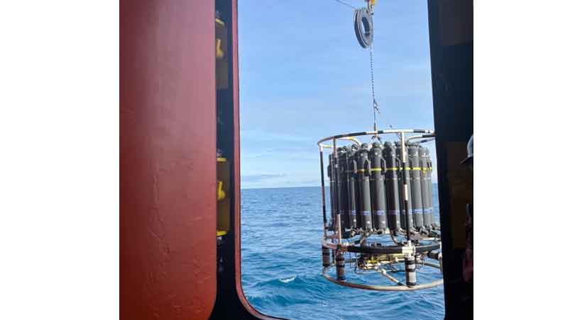
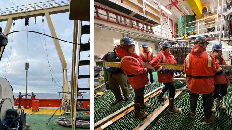
CTD rosette being lowered into the water off the side of the R/V Nathaniel B. Palmer.
Exciting news from the R/V Nathaniel B. Palmer!
We have officially collected our first samples. This is a momentous occasion as it puts our team one step closer to addressing our research questions and better understanding microbial communities and their influence on methane hydrates.
With this goal in mind – we sampled the sediment and water column. While the sediment is where the bulk of our samples will be collected from, understanding aspects of the water column will play an important role in this project.
First, we had our CTD cast to survey the water column. This sampling process is akin to filling a bottle of seawater at various depths. The CTD will measure conductivity, temperature, and depth (CTD) while collecting water samples from several depths in the water column in a single deployment. A CTD rosette (Figure 1), gets lowered by cranes into the water and travels to the bottom of the water column to start the sampling process. As the CTD moves from deep to shallow, onboard the R/V Palmer, technicians will close bottles at each depth. Once the CTD gets to the surface, we have an assortment of water samples ranging from 5 to 570 meters.
The CTD is then brought back to the ship where thirsty researchers are ready to divide and conquer. The order of sub-sampling and the amount each research team is able to collect is usually negotiated ahead of time – ensuring each group has enough water for their research. Working as a team with the Maiti Lab (Louisiana State University), Coffin Lab (Texas A&M University–Corpus Christi), and the Jeffrey lab (University of West Florida), water samples were quickly subsampled.
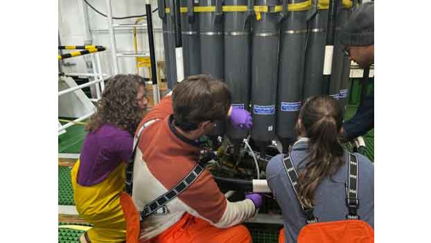
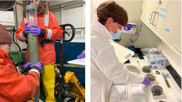
Dr. Brandi Kiel Reese, Caleb Boyd, Hannah Organ (Texas A&M University-Corpus Christi), and Dr. Kanchan Maiti (Louisiana State University) taking water samples from the CTD.
Caleb Boyd and Hannah Organ then took our samples and began filtering. These filter sizes were targeting the microbial community and lipids.
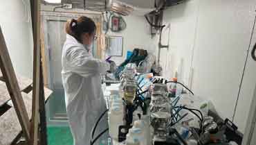
Hannah Organ (Texas A&M University Corpus Christi) filtering water samples.
While filtering, a multicore was lowered passing through the water column and entering the ocean floor. The multicore has the ability to take 12 sediment cores simultaneously. Each core is roughly 30-40 cm in length and is clear on the outside. This allows us to see the color change in the sediment column (as well as check for any critters that might have been captured) (Figure 4). These cores are then capped remotely and brought back to the deck of the R/V Palmer.
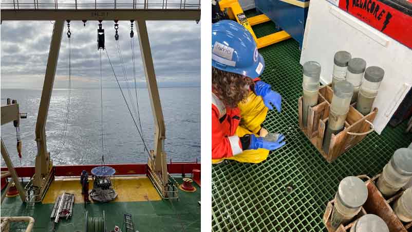
A) Multicore ready to be deployed to sample sediment. B) Dr. Brandi Kiel Reese investigating the retrieved sediment cores.
Our team of researchers then got to work sub sectioning each core. We first remove any overlying water and then place the core on an extruder stand. This pushes the sediment out to the top where we can then slice off measured sections of sediment that look like slices of cake. During this process we note the characteristics of the sediment.
A) Dr. Brandi Kiel Reese and Rachel Weisend preparing to section the sediment core by placing it on the extruder, B) Caleb Boyd with sectioned sediment prepping for cell counts and preservation.
- Does it smell organic? Or like rotten eggs?
- Does the sediment fall apart easily? Or does it stick together?
- Is the sediment more blue in color or is it a brown red color?
Describing the sediment visually starts to clue us in on the types of microbes that we may find. Post sub sectioning our samples make their way into tubes for cell counts and community analysis, and into bags for preservation in our -80 C freezer.
By the time we finished sampling our multicore samples, we were called to help carry the 3 meter long Kasten core into the lab (Figure 6).
A) 3 meter Kasten core being lowered onto the deck, B) Rachel Weisend and Alyssa Cotton (University of West Florida) helping carry the Kasten core into the lab.
From here, geologists from the Prothro lab (Texas A&M University Corpus Christi) and Bart Lab (Louisiana State University), begin describing characteristics in the sediment such as grain size and color, as well as any rapid changes between the two. We were able to quickly grab 40 grams of sediment from various points within the core for community analyses.
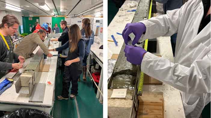
A) Dr. Phillip Bart (Louisiana State University), Dr. Lindsey Prothro Kaple (Texas A&M University – Corpus Christi) and their teams prepping the Kasten core, B) Caleb Boyd taking a sediment sample from the Kasten core.
This entire day took roughly 18 hours from morning preparations to clean up and celebrated a job well done by getting some well deserved rest. From here we will continue our transit to McMurdo Station and daydream of processing our samples back on land.
More updates to come!
Written by Rachel Weisend
(-075 28.201 S, -179 19.954 W)